Neuropsychological Deficits After Concussion Are Correlated with White Matter Abnormalities
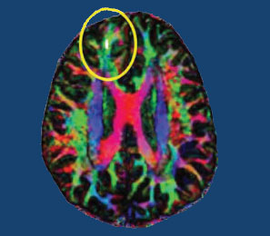
Many people suffer problems with mental functioning after an apparent concussion (otherwise known as mild traumatic brain injury, or mTBI) that does not show abnormalities on traditional brain imaging measures such as the MRI. New technology called diffusion tensor imaging (DTI) shows that the integrity of white matter tracts may be disturbed by concussions. White matter comprises parts of the brain where myelin wraps around axons, as opposed to grey matter, which reflects the presence of neuronal cell bodies.
In a longitudinal study published in the Journal of Neurotrauma, Vigneswaran Veeramuthu and colleagues compared 61 people with an mTBI to 19 healthy controls. The mTBI participants had their neuropsychological faculties assessed an average of 4.35 hours after their trauma, and participated in DTI scans an average of 10 hours after the trauma. Both the neuropsychological assessment and the DTI scan were repeated six months later. When the acute and follow-up assessments were compared to the same assessments in control participants, the two groups showed differences in numerous white matter tracts at the six-month mark. There was also an association between the degree of abnormality observed on the DTI scans and decrements in performance on the tests of neuropsychological functioning both immediately after the trauma and six months later.
The researchers concluded that their results “provide new evidence for the use of DTI as an imaging biomarker and indicator of [white matter] damage occurring in the context of mTBI, and [the results] underscore the dynamic nature of brain injury and possible biological basis of chronic neurocognitive alterations.”